. 2016 Mar;124(3):706-20.
doi: 10.1097/ALN.0000000000000993.

Activation of Peripheral μ-opioid Receptors by Dermorphin [D-Arg2, Lys4] (1-4) Amide Leads to Modality-preferred Inhibition of Neuropathic Pain

Affiliations

Activation of Peripheral μ-opioid Receptors by Dermorphin [D-Arg2, Lys4] (1-4) Amide Leads to Modality-preferred Inhibition of Neuropathic Pain

Vinod Tiwari et al. Anesthesiology. 2016 Mar.

Abstract

Background: Opioids have long been regarded as the most effective drugs for the treatment of severe acute and chronic pain. Unfortunately, their therapeutic efficacy and clinical utility have been limited because of central and peripheral side effects.

Methods: To determine the therapeutic value of peripheral μ-opioid receptors as a target for neuropathic pain treatment, the authors examined the effects of dermorphin [D-Arg2, Lys4] (1-4) amide (DALDA), a hydrophilic, peripherally acting μ-opioid receptor agonist, in male and female rats with spinal nerve ligation-induced neuropathic pain. The authors also utilized behavioral, pharmacologic, electrophysiologic, and molecular biologic tools to characterize DALDA's possible mechanisms of action in male rats.

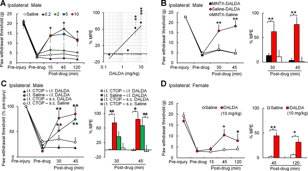
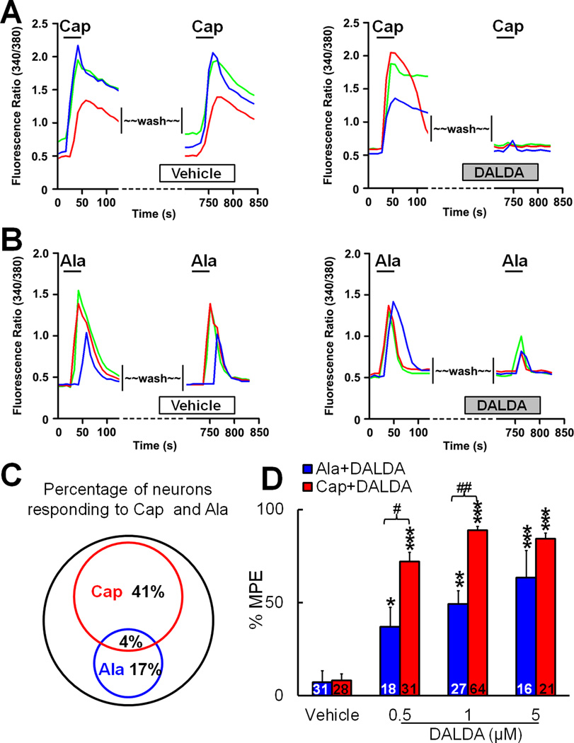
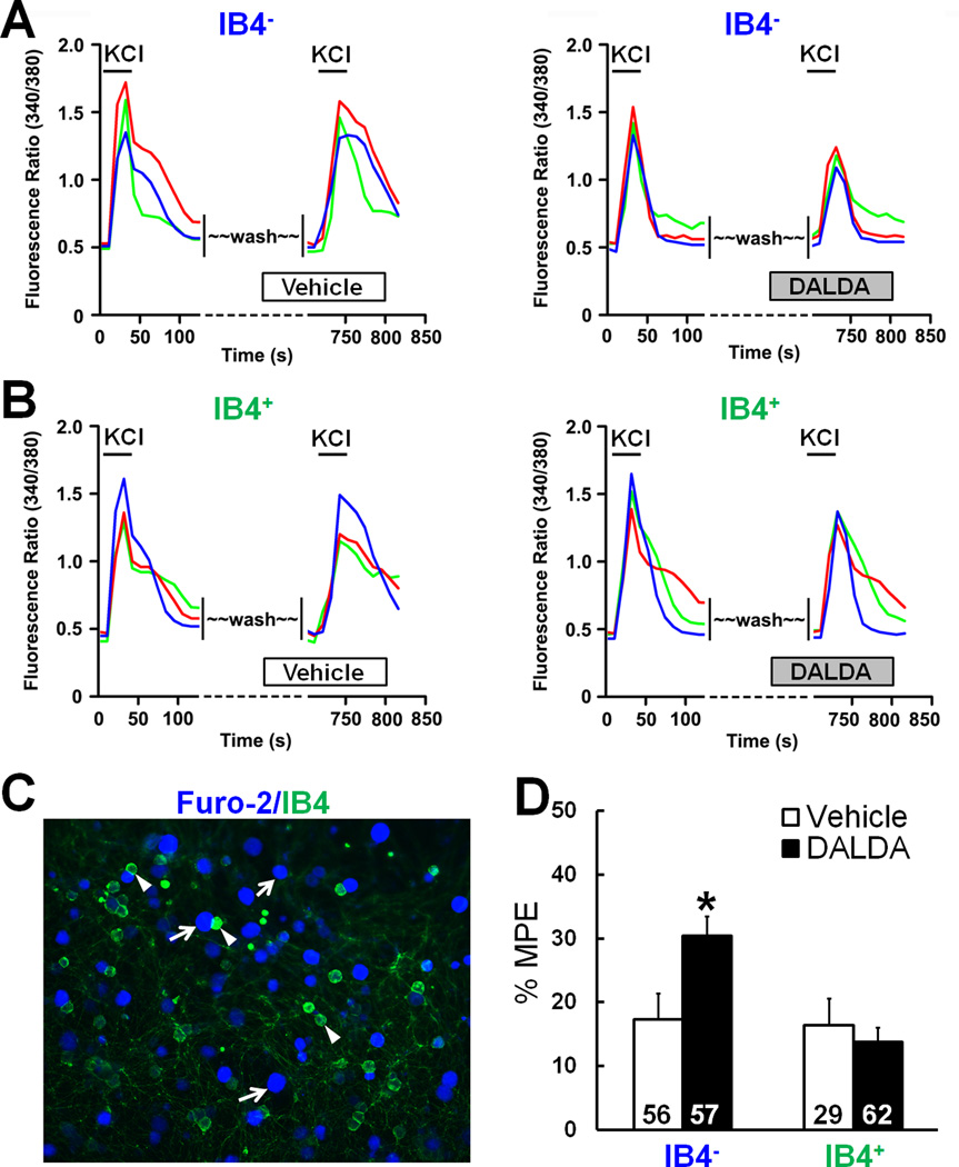
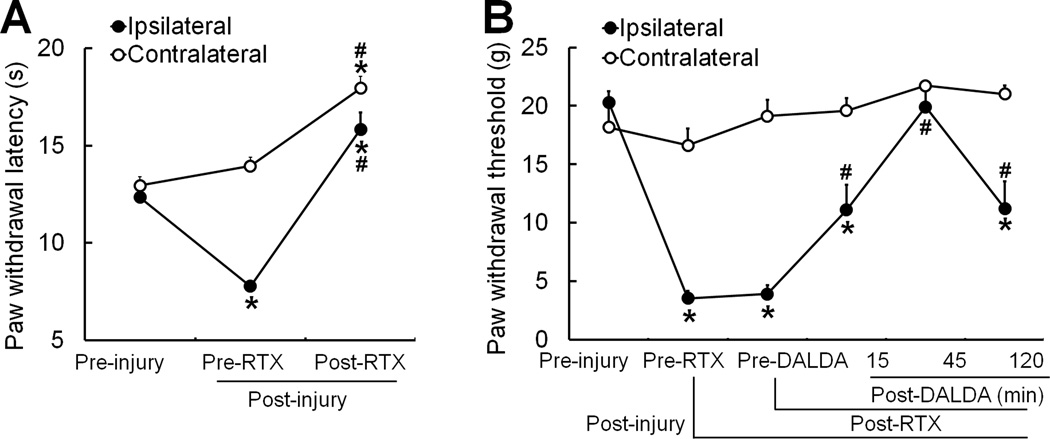
Results: DALDA, administered subcutaneously, had 70 times greater efficacy for inhibiting thermal (n = 8 to 11/group) than mechanical hypersensitivity (n = 6 to 8/group) in male rats. The pain inhibitory effects of DALDA on mechanical and heat hypersensitivity were abolished in animals pretreated with systemic methylnaltrexone (n = 7 to 9/group), a peripheral μ-opioid receptor antagonist. In the spinal wide-dynamic range neurons, systemic DALDA inhibited C-fiber-mediated, but not A-fiber-mediated, response in neuropathic male rats (n = 13). In primary sensory neurons, DALDA inhibited the capsaicin-induced [Ca2+] increase more than the β-alanine-induced [Ca] increase (n = 300); capsaicin and β-alanine activate subpopulations of neurons involved in the signaling of heat and mechanical pain, respectively. DALDA-treated rats (n = 5 to 8/group) did not exhibit motor deficits and locomotor impairment suggesting that it does not induce central side effects.

Conclusions: These findings suggest that DALDA may represent a potential alternative to current opioid therapy for the treatment of neuropathic pain and is likely to be associated with minimal adverse effects.


Figures

Figure 1
Figure 1. Systemic administration of dermorphin [D-Arg2, Lys4] (1–4) amide (DALDA) inhibits mechanical hypersensitivity in nerve-injured male rats through activation of peripheral opioid receptors
(A) Left: Subcutaneous (s.c.) injection of DALDA (0.2, 2, 5, 10 mg/kg, n=6–8/dose) dose-dependently inhibited mechanical allodynia in male rats at 2–3 weeks after spinal nerve ligation (SNL), as indicated by the significant increase in ipsilateral paw withdrawal threshold (PWT). The time course shows peak drug effect at 45 minutes after injection. *p<0.05 versus pre-drug (5, 10mg/kg), two-way mixed model ANOVA. Right: The maximum possible effect (MPE) of DALDA at 45 minutes post-drug was calculated. MPE (%) = [1 − (Pre-injury PWT − Post-drug PWT)]/[Pre-injury PWT − Pre-drug PWT] × 100. **p<0.01, ***p<0.001 versus saline group (n=11), one-way ANOVA. (B) Left: Pretreatment with an intraperitoneal (i.p.) injection of methylnaltrexone (MNTX, 5 mg/kg, 10-minute pretreatment, n=7), but not saline (n=7), blocked the inhibitory effect of DALDA (10 mg/kg, s.c.) on mechanical hypersensitivity in male SNL rats. Injection of methylnaltrexone (5 mg/kg, i.p., n=7) followed by saline (s.c.) did not change PWT from pre-drug baseline. Right: The MPEs at 30 minutes and 45 minutes after the second drug administration were calculated for each group. (C) Left: Intrathecal (i.th.) pretreatment with the highly selective MOR antagonist D-Phe-Cys-Tyr-D-Trp-Orn-Thr-Pen-Thr-NH2 (CTOP, 5 µg/10 µl, n=5), but not saline (n=5), blocked inhibition of mechanical hypersensitivity by intrathecal DALDA (0.5 µg/10 µl) in male SNL rats. However, the same CTOP treatment did not block inhibition of mechanical hypersensitivity by systemic DALDA (10 mg/kg, s.c., n=7). Intrathecal CTOP (5 µg/10 µl, n=5) followed by saline injection (s.c.) did not change PWT from pre-drug baseline. (D) Left: DALDA (10 mg/kg, n=6, s.c.), but not saline (n=6), also inhibited mechanical allodynia in female rats at 2–3 weeks after SNL. Right: The MPE at 45 and 120 minutes post-drug was calculated. (B–D) Left: *p<0.05, **p<0.01 versus pre-drug, two-way mixed model ANOVA. Right: *p<0.05, **p<0.01 versus saline, one-way ANOVA. Data are expressed as mean ± SEM.
Figure 2
Figure 2. Systemic administration of dermorphin [D-Arg2, Lys4] (1–4) amide (DALDA) inhibits heat hypersensitivity in nerve-injured male rats at lower doses than those required to inhibit mechanical hypersensitivity
(A) At 2–3 weeks after spinal nerve ligation (SNL) in male rats, subcutaneous (s.c.) injection of DALDA (0.02, 0.1, 0.2, 2 mg/kg, n=8–11/group) dose-dependently increased the ipsilateral paw withdrawal latency (PWL) as compared to that at pre-drug baseline. *p<0.05 versus pre-drug (0.1. 0.2, 2 mg/kg), two-way mixed model ANOVA. (B) The PWL in the contralateral hindpaw did not change after nerve injury or drug treatment. (C) The maximum possible effect (MPE) of DALDA at 45 minutes post-drug was calculated. MPE (%) = [1 − (Pre-injury PWL − Post-drug PWL)/(Pre-injury PWL − Pre-drug PWL)] × 100. **p<0.01, ***p<0.001 versus saline group (n=11), one-way ANOVA. (D) Pretreatment with an intraperitoneal (i.p.) injection of methylnaltrexone (MNTX, 5 mg/kg, n=9), but not saline (n=9), 10 minutes before DALDA (2 mg/kg, s.c.) blocked inhibition of heat hyperalgesia by DALDA in male rats. Methylnaltrexone (5 mg/kg) followed by saline (s.c.) did not change PWL from pre-drug baseline (n=9). *p<0.05 versus pre-drug, #p<0.05 versus MNTX+DALDA group, two-way mixed model ANOVA. (E) The PWL of the contralateral hindpaw did not change after nerve injury or drug treatment. (F–G) DALDA (10 mg/kg, n=6, s.c.), but not saline (n=6), also inhibited heat hypersensitivity in female rats at 2–3 weeks after SNL. *p<0.05 versus pre-drug, #p<0.05 versus saline, two-way mixed model ANOVA. Data are expressed as mean ± SEM.
Figure 3
Figure 3. Systemic dermorphin [D-Arg2, Lys4] (1–4) amide (DALDA) selectively inhibits the C-component of WDR neurons to electrical stimulation, an effect that is blocked by methylnaltrexone
(A) An analog recording of wide-dynamic range (WDR) neuronal responses to the 1st, 4th, 8th, and 16th stimulus of a train of intracutaneous electrical stimuli (0.5 Hz, 16 pulses, 2.0 milliseconds, supra-C-fiber activation threshold) that induces windup. WDR neuronal responses display A- and C-components to an intracutaneous electrical stimulus. At 2–3 weeks after spinal nerve ligation (SNL) in male rats, windup of C-component was inhibited at 30–45 minutes after systemic administration of DALDA (10 mg/kg, i.p.). (B) The total A-component and C-component to graded intracutaneous electrical stimuli (0.1–10 mA, 2.0 milliseconds) and the total C-component to windup-inducing stimuli after treatment of SNL rats with saline (i.p., n=10), DALDA (10 mg/kg, i.p., n=13), and methylnaltrexone (MNTX, 5 mg/kg, i.p., 15-minute pretreatment, n=9) followed by DALDA (10 mg/kg). **p<0.01 versus saline group, one-way ANOVA. (C) The stimulus-response (S-R) function of the A-component of WDR neuronal response to graded intracutaneous electrical stimuli (0.1–10 mA, 2 milliseconds) before and 30–45 minutes after systemic injection of DALDA (upper panel, 10 mg/kg, n=9, i.p.) or methylnaltrexone (MNTX, 5 mg/kg, i.p., 15-minute pretreatment) with DALDA (lower panel, 10 mg/kg, n=8, i.p.). (D) The S-R function of C-component to graded intracutaneous electrical stimuli in each group. (E) Windup of C-component of WDR neurons to a train of intracutaneous electrical stimuli (0.5 Hz, 16 pulses) before and after drug treatment. The C-component to 0.5 Hz stimulation was plotted against the stimulation sequence number of each trial. C-E: *p<0.05 versus pre-drug, two-way repeated measures ANOVA. Data are expressed as mean ± SEM.
Figure 4
Figure 4. Effects of systemic dermorphin [D-Arg2, Lys4] (1–4) amide (DALDA) on the responses of wide-dynamic range (WDR) neurons to mechanical stimulation in nerve-injured rats
(A) Left: Peri-stimulus time histograms (bin size: 0.2 seconds) show an example of WDR neuronal response to punctuate mechanical stimuli (1.0–15.0 g von Frey probe, 5 seconds, applied to the skin receptive field) that did not change after subcutaneous (s.c.) injection of DALDA (10 mg/kg, non-responding). Right: An example of another WDR neuron that shows decreased response after DALDA treatment (responding). (B) At 2–3 weeks after spinal nerve ligation (SNL) in male rats, the response to brushing stimuli and the stimulus-response functions of WDR neuronal response to graded mechanical stimuli were not significantly different from pre-drug baseline at 30–45 minutes after DALDA injection (10 mg/kg, s.c., n=13). (C) Injection of saline did not change WDR neuronal response to mechanical stimuli in SNL rats (n=8). (D) The total response of each WDR neuron to graded mechanical stimuli was plotted for each group (as % pre-drug value). Inhibitory cells were defined as neurons that had a post-drug response that was <74% of pre-drug level (below red dashed line), which is more than two SD less than the mean of the saline group (122 ± 24%, mean ± SD, n=8). The facilitatory cells were defined as neurons that had a post-drug response >170% of pre-drug level (above blue dashed line). Black bar: mean response. Data are expressed as mean + SEM. APs, action potentials.
Figure 5
Figure 5. Dermorphin [D-Arg2, Lys4] (1–4) amide (DALDA) inhibits capsaicin- and β-alanine–induced increases in [Ca2+] in dorsal root ganglion (DRG) neurons
(A) Left: Representative traces from calcium-imaging assays show the increase of [Ca2+] in cultured DRG neurons in response to the first and second bath application of capsaicin (Cap, 0.5 µM). Neurons were washed (10 minutes) with vehicle before the second Cap application. Right: Pretreatment with DALDA (1 µM, 10 minutes, bath application) blocked the increase in [Ca2+] induced by the second application of Cap. (B) Left: Representative traces from calcium-imaging assays show the increase of [Ca2+] in cultured DRG neurons in response to the first and second bath application of β-alanine (Ala, 1 mM). Neurons were washed with vehicle before the second Ala application. Right: Pretreatment with DALDA (1 µM, 10 minutes) partially reduced the increase in [Ca2+] induced by the second application of Ala. (C) The Venn diagram of calcium responses illustrates the proportion of Cap- and Ala-responsive DRG neurons (n=300). The sizes of the circles are proportional to the sizes of the cell populations. (D) Quantification of calcium-imaging assays. DALDA (0.5, 1, 5 µM) dose-dependently inhibited Cap- and Ala-induced increases in [Ca2+]. MPE (%) = [(Pre-DALDA) − (Post-DALDA)]/(Pre-DALDA)] × 100. Numbers of neurons in each group are indicated. *p<0.05, **p<0.01,***p<0.001 versus vehicle group; #p<0.05, ##p<0.01 versus Ala-DALDA group, one-way ANOVA. Data are expressed as mean + SEM.
Figure 6
Figure 6. Dermorphin [D-Arg2, Lys4] (1–4) amide (DALDA) inhibits potassium chloride (KCl)-induced increases in [Ca2+] in isolectin IB4 dorsal root ganglion (DRG) neurons
(A) Left: Representative traces from calcium-imaging assays show the increase of [Ca2+] in isolectin IB4 DRG neurons after the first and second bath application of KCl (30 mM). Neurons were washed with vehicle (10 minutes) before the second KCl application. Right: The [Ca2+] increase in response to the second application of KCl was reduced in IB4 neurons by pretreatment with DALDA (1 µM, 10 minutes, bath application). (B) DALDA did not reduce the KCl-evoked increase in [Ca2+] in IB4+ neurons. (C) Fluorescent image of DRG neurons after they were labeled with IB4-FITC and loaded with Fura 2-acetomethoxyl ester. Examples of IB4 (purple) and IB4+ (green) neurons are marked with arrows and arrowheads, respectively. (D) Quantification of calcium-imaging assays. DALDA significantly inhibited the KCl-induced increase in [Ca2+] in IB4 neurons, but not in IB4+ neurons. MPE (%) = [(Pre-DALDA) − (Post-DALDA)]/(Pre-DALDA)] × 100. Data are expressed as mean + SEM. *p<0.05 versus vehicle group, Student's t-test.
Figure 7
Figure 7. Systemic dermorphin [D-Arg2, Lys4] (1–4) amide (DALDA) retains ability to inhibit mechanical hypersensitivity in nerve-injured rats that receive resiniferatoxin (RTX)
(A) The paw withdrawal latency (PWL) of the ipsilateral hindpaw was significantly decreased in male rats on day 7 after spinal nerve ligation (SNL, pre-RTX). On days 7–9 after intraperitoneal (i.p.) injection of RTX (0.1 mg/kg, n=9), both ipsilateral and contralateral PWLs were significantly increased from the pre-RTX and pre-injury level. (B) Rats showed significant reduction in ipsilateral paw withdrawal thresholds (PWTs) on day 7 post-SNL. Systemic RTX treatment (0.1 mg/kg, i.p., n=9) did not alter SNL-induced mechanical allodynia. However, systemic administration of DALDA (10 mg/kg, s.c.) significantly attenuated mechanical allodynia even after RTX treatment. *p<0.05 versus pre-injury, #p<0.05 versus pre-RTX, one-way repeated measures ANOVA. Data are expressed as mean + SEM.
Figure 8
Figure 8. Systemic dermorphin [D-Arg2, Lys4] (1–4) amide (DALDA) does not induce opioid-related side effects and does not affect exploration activity of rats
(A) In the rota-rod test, neither DALDA (10 mg/kg, s.c., n=5) nor saline (n=8) decreased fall time (i.e., time on the rod) in spinal nerve ligation (SNL) male rats at 45 minutes post-injection, as compared to that at baseline. (B) Morphine (10 mg/kg, s.c., n=8), but not a higher dose of DALDA (20 mg/kg, n=8), did induce motor dysfunction on the rota-rod test at 45 minutes post-injection. (C) Examples of SNL rat exploration activity (10 minutes' duration) in the open field test before and 45 minutes after injection of saline, DALDA (10 mg/kg, s.c.), and morphine (10 mg/kg, s.c., n=5/group). (D–E) In the open field test, morphine (10 mg/kg, s.c.), but not DALDA (10 mg/kg, s.c.) or saline (n=5/group), reduced the total distance traveled in 10 minutes and the number of center crossings by SNL rats at 45 minutes after injection. *p<0.05, **p<0.01 versus pretreatment, paired t-test. Data are expressed as mean + SEM.

Similar articles

Cited by

References

    1. Mathieson S, Maher CG, Terwee CB, Folly de CT, Lin CW. Neuropathic pain screening questionnaires have limited measurement properties. A systematic review. J. Clin. Epidemiol. 2015;68(8):957–966. - PubMed
    1. Attal N, Bouhassira D. Pharmacotherapy of neuropathic pain: which drugs, which treatment algorithms? Pain. 2015;156(Suppl 1):S104–S114. - PubMed
    1. Gewandter JS, Dworkin RH, Turk DC, Farrar JT, Fillingim RB, Gilron I, Markman JD, Oaklander AL, Polydefkis MJ, Raja SN, Robinson JP, Woolf CJ, Ziegler D, Ashburn MA, Burke LB, Cowan P, George SZ, Goli V, Graff OX, Iyengar S, Jay GW, Katz J, Kehlet H, Kitt RA, Kopecky EA, Malamut R, McDermott MP, Palmer P, Rappaport BA, Rauschkolb C, Steigerwald I, Tobias J, Walco GA. Research design considerations for chronic pain prevention clinical trials: IMMPACT recommendations. Pain. 2015;156(7):1184–1197. - PMC - PubMed
    1. Campbell JN, Meyer RA. Mechanisms of neuropathic pain. Neuron. 2006;52:77–92. - PMC - PubMed
    1. Kim KJ, Yoon YW, Chung JM. Comparison of three rodent neuropathic pain models. Exp. Brain Res. 1997;113:200–206. - PubMed

Publication types