

Kidney Disease Progression Does Not Decrease Intestinal Phosphorus Absorption in a Rat Model of Chronic Kidney Disease-Mineral Bone Disorder
- PMID: 31618470
- PMCID: PMC7012714
- DOI: 10.1002/jbmr.3894
Kidney Disease Progression Does Not Decrease Intestinal Phosphorus Absorption in a Rat Model of Chronic Kidney Disease-Mineral Bone Disorder
Abstract
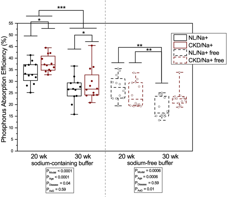
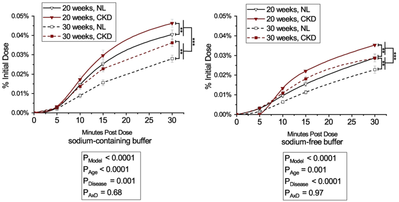
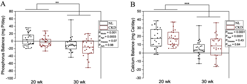
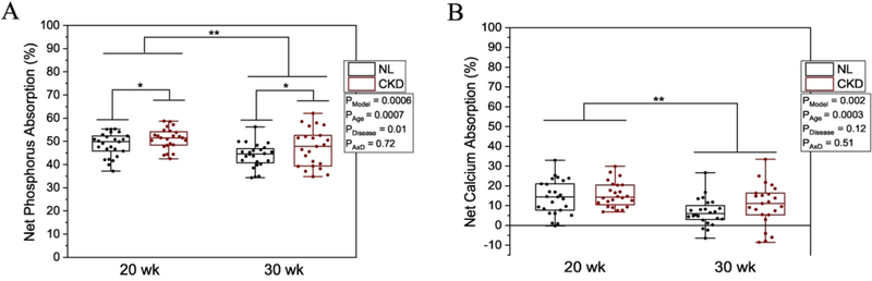
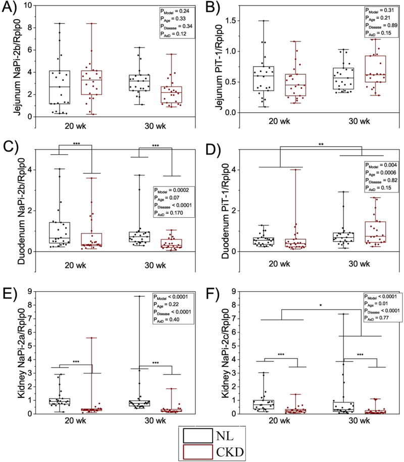
The Cy/+ rat has been characterized as a progressive model of chronic kidney disease-mineral bone disorder (CKD-MBD). We aimed to determine the effect of kidney disease progression on intestinal phosphorus absorption and whole-body phosphorus balance in this model. A total of 48 Cy/+ (CKD) and 48 normal littermates (NL) rats were studied at two ages: 20 weeks and 30 weeks, to model progressive kidney function decline at approximately 50% and 20% of normal kidney function. Sodium-dependent and sodium-independent intestinal phosphorus absorption efficiency were measured by the in situ jejunal ligated loop method using 33 P radioisotope. Our results show that CKD rats had slightly higher sodium-dependent phosphorus absorption compared to NL rats, and absorption decreased from 20 to 30 weeks. These results are in contrast to plasma 1,25OH2 D, which was lower in CKD rats. Gene expression of the major intestinal phosphorus transporter, NaPi-2b, was not different between CKD and NL rats in the jejunum but was lower in CKD rats versus NL rats in the duodenum. Jejunal ligated loop phosphorus absorption results are consistent with percent net phosphorus absorption results obtained from metabolic balance: higher net percent phosphorus absorption values in CKD rats compared with NL, and lower values in 30-week-olds compared with 20-week-olds. Phosphorus balance was negative (below zero) in CKD rats, significantly lower in 30-week-old rats compared with 20-week-old rats, and lower in CKD rats compared with NL rats at both ages. These results demonstrate no reduction in intestinal phosphorus absorption with progression of CKD despite lower 1,25OH2 D status when assessed by an in situ ligated loop test, which is in contrast to the majority of in vitro studies, and if confirmed in further studies, could challenge the physiological relevance of in vitro findings. © 2019 American Society for Bone and Mineral Research.
Keywords: ANIMAL MODELS; DISORDERS OF CALCIUM/PHOSPHATE METABOLISM; GENETIC ANIMAL MODELS; NUTRITION; PTH/VIT D/FGF23.
© 2019 American Society for Bone and Mineral Research.
Figures
Similar articles
-
The pathophysiology of early-stage chronic kidney disease-mineral bone disorder (CKD-MBD) and response to phosphate binders in the rat.J Bone Miner Res. 2011 Nov;26(11):2672-81. doi: 10.1002/jbmr.485. J Bone Miner Res. 2011. PMID: 21826734
-
Effects of ferric citrate and intravenous iron sucrose on markers of mineral, bone, and iron homeostasis in a rat model of CKD-MBD.Nephrol Dial Transplant. 2022 Sep 22;37(10):1857-1867. doi: 10.1093/ndt/gfac162. Nephrol Dial Transplant. 2022. PMID: 35482713 Free PMC article.
-
Effect of dietary phosphorus intake and age on intestinal phosphorus absorption efficiency and phosphorus balance in male rats.PLoS One. 2018 Nov 19;13(11):e0207601. doi: 10.1371/journal.pone.0207601. eCollection 2018. PLoS One. 2018. PMID: 30452474 Free PMC article.
-
Intestinal Phosphorus Absorption in Chronic Kidney Disease.Nutrients. 2018 Sep 23;10(10):1364. doi: 10.3390/nu10101364. Nutrients. 2018. PMID: 30249044 Free PMC article. Review.
-
Oh, My Gut! New insights on the role of the gastrointestinal tract and the gut microbiome in chronic kidney disease-mineral and bone disorder.Curr Opin Nephrol Hypertens. 2024 Mar 1;33(2):226-230. doi: 10.1097/MNH.0000000000000961. Epub 2023 Dec 13. Curr Opin Nephrol Hypertens. 2024. PMID: 38088374 Free PMC article. Review.
Cited by
-
Phosphate Balance and CKD-Mineral Bone Disease.Kidney Int Rep. 2021 May 17;6(8):2049-2058. doi: 10.1016/j.ekir.2021.05.012. eCollection 2021 Aug. Kidney Int Rep. 2021. PMID: 34386654 Free PMC article. Review.
-
Acute High Dietary Phosphorus Following Low-Phosphorus Diet Acclimation Does Not Enhance Intestinal Fractional Phosphorus Absorption in Nephrectomized Male Rats.JBMR Plus. 2022 Nov 16;6(12):e10698. doi: 10.1002/jbm4.10698. eCollection 2022 Dec. JBMR Plus. 2022. PMID: 36530183 Free PMC article.
-
Adverse Effects of Autoclaved Diets on the Progression of Chronic Kidney Disease and Chronic Kidney Disease-Mineral Bone Disorder in Rats.Am J Nephrol. 2020;51(5):381-389. doi: 10.1159/000506729. Epub 2020 Mar 6. Am J Nephrol. 2020. PMID: 32146472 Free PMC article.
-
Effect of nutritional calcium and phosphate loading on calciprotein particle kinetics in adults with normal and impaired kidney function.Sci Rep. 2022 May 5;12(1):7358. doi: 10.1038/s41598-022-11065-3. Sci Rep. 2022. PMID: 35513558 Free PMC article.
-
Feeling gutted in chronic kidney disease (CKD): Gastrointestinal disorders and therapies to improve gastrointestinal health in individuals CKD, including those undergoing dialysis.Semin Dial. 2024 Jul-Aug;37(4):334-349. doi: 10.1111/sdi.13030. Epub 2021 Oct 27. Semin Dial. 2024. PMID: 34708456 Free PMC article. Review.
References
-
- Moe S, Drueke T, Cunningham J, Goodman W, Martin K, Olgaard K, et al. Definition, evaluation, and classification of renal osteodystrophy: a position statement from Kidney Disease: Improving Global Outcomes (KDIGO). Kidney Int 2006;69(11):1945–53. - PubMed
-
- Levin A, Bakris GL, Molitch M, Smulders M, Tian J, Williams LA, et al. Prevalence of abnormal serum vitamin D, PTH, calcium, and phosphorus in patients with chronic kidney disease: results of the study to evaluate early kidney disease. Kidney Int. 2007;71(1):31–8. - PubMed
-
- Gansevoort RT, Correa-Rotter R, Hemmelgarn BR, Jafar TH, Heerspink HJ, Mann JF, et al. Chronic kidney disease and cardiovascular risk: epidemiology, mechanisms, and prevention. Lancet. 2013;382(9889):339–52. - PubMed
Publication types
MeSH terms
Substances
Grants and funding
LinkOut - more resources
Full Text Sources
Medical
Research Materials
