

SCN1B-linked early infantile developmental and epileptic encephalopathy
- PMID: 31709768
- PMCID: PMC6917350
- DOI: 10.1002/acn3.50921
SCN1B-linked early infantile developmental and epileptic encephalopathy
Abstract
Objective: Patients with Early Infantile Epileptic Encephalopathy (EIEE) 52 have inherited, homozygous variants in the gene SCN1B, encoding the voltage-gated sodium channel (VGSC) β1 and β1B non-pore-forming subunits.
Methods: Here, we describe the detailed electroclinical features of a biallelic SCN1B patient with a previously unreported variant, p.Arg85Cys.
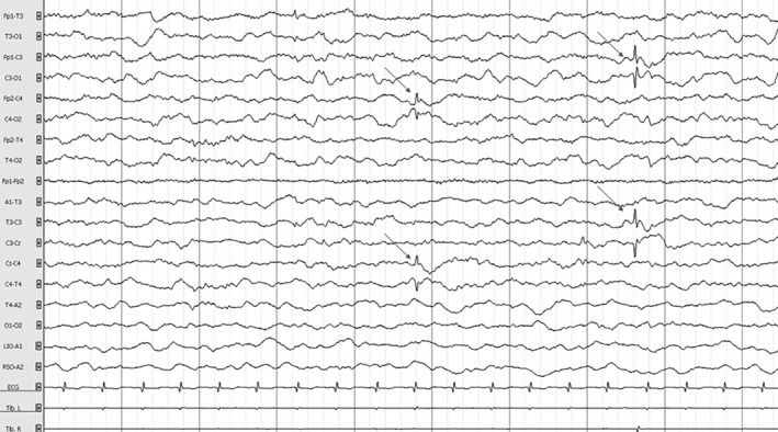
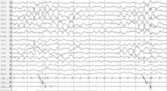
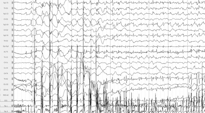
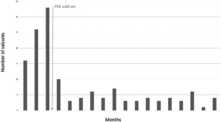
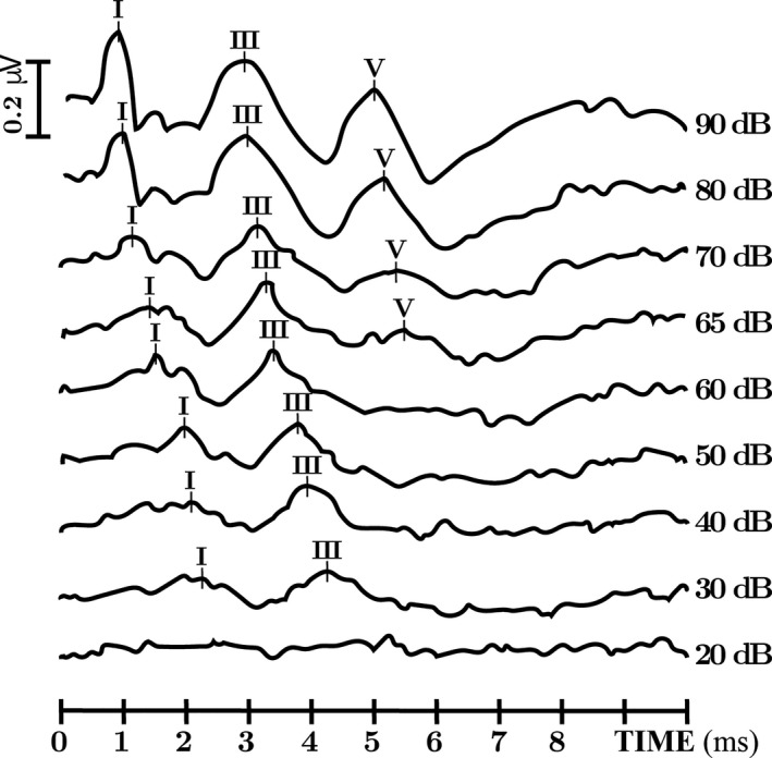
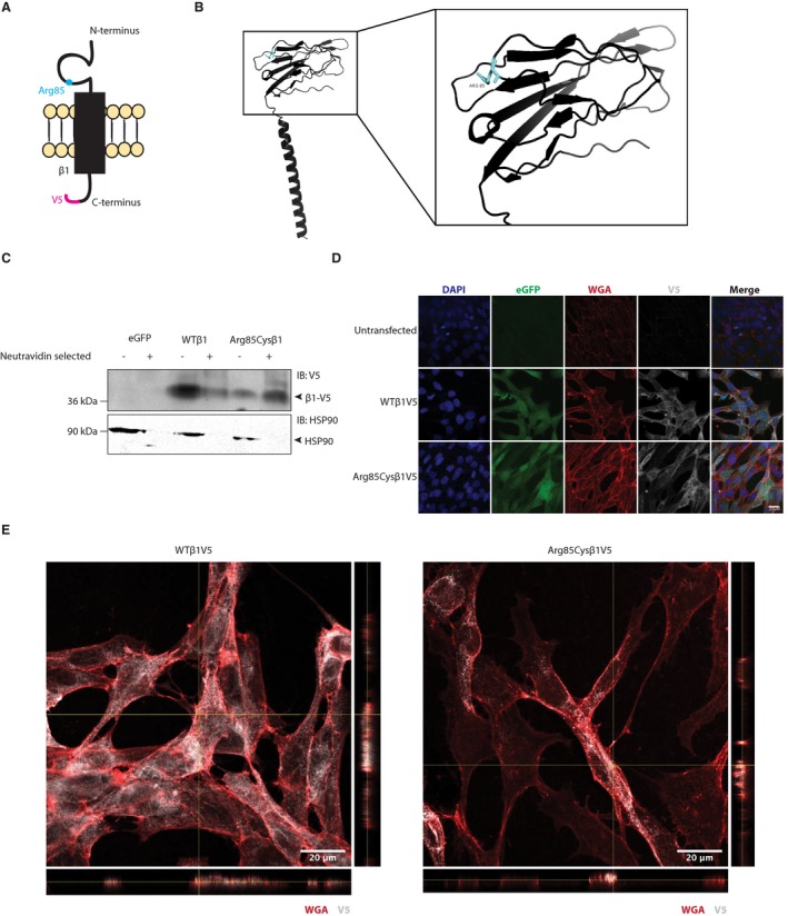
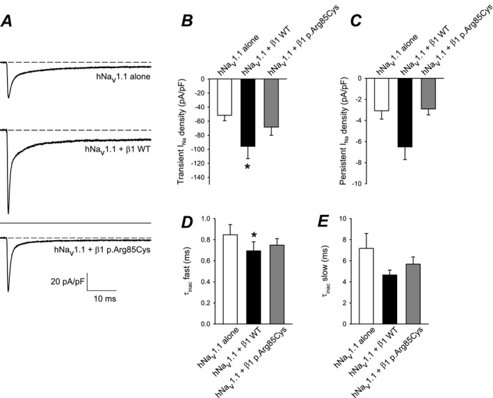
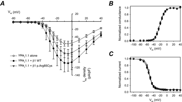
Results: The female proband showed hypotonia from birth, multifocal myoclonus at 2.5 months, then focal seizures and myoclonic status epilepticus (SE) at 3 months, triggered by fever. Auditory brainstem response (ABR) showed bilateral hearing loss. Epilepsy was refractory and the patient had virtually no development. Administration of fenfluramine resulted in a significant reduction in seizure frequency and resolution of SE episodes that persisted after a 2-year follow-up. The patient phenotype is more compatible with early infantile developmental and epileptic encephalopathy (DEE) than with typical Dravet syndrome (DS), as previously diagnosed for other patients with homozygous SCN1B variants. Biochemical and electrophysiological analyses of the SCN1B variant expressed in heterologous cells showed cell surface expression of the mutant β1 subunit, similar to wild-type (WT), but with loss of normal β1-mediated modification of human Nav 1.1-generated sodium current, suggesting that SCN1B-p.Arg85Cys is a loss-of-function (LOF) variant.
Interpretation: Importantly, a review of the literature in light of our results suggests that the term, early infantile developmental and epileptic encephalopathy, is more appropriate than either EIEE or DS to describe biallelic SCN1B patients.
© 2019 The Authors. Annals of Clinical and Translational Neurology published by Wiley Periodicals, Inc on behalf of American Neurological Association.
Conflict of interest statement
The authors declare no conflict of interest.
Figures

Similar articles
-
Homozygous SCN1B variants causing early infantile epileptic encephalopathy 52 affect voltage-gated sodium channel function.Epilepsia. 2021 Jun;62(6):e82-e87. doi: 10.1111/epi.16913. Epub 2021 Apr 26. Epilepsia. 2021. PMID: 33901312 Free PMC article.
-
Excitatory and inhibitory neuron defects in a mouse model of Scn1b-linked EIEE52.Ann Clin Transl Neurol. 2020 Nov;7(11):2137-2149. doi: 10.1002/acn3.51205. Epub 2020 Sep 26. Ann Clin Transl Neurol. 2020. PMID: 32979291 Free PMC article.
-
Developmental and epileptic encephalopathy in two siblings with a novel, homozygous missense variant in SCN1B.Am J Med Genet A. 2019 Nov;179(11):2190-2195. doi: 10.1002/ajmg.a.61344. Epub 2019 Aug 29. Am J Med Genet A. 2019. PMID: 31465153
-
SCN1B Genetic Variants: A Review of the Spectrum of Clinical Phenotypes and a Report of Early Myoclonic Encephalopathy.Children (Basel). 2022 Oct 1;9(10):1507. doi: 10.3390/children9101507. Children (Basel). 2022. PMID: 36291443 Free PMC article. Review.
-
New focus on cardiac voltage-gated sodium channel β1 and β1B: Novel targets for treating and understanding arrhythmias?Heart Rhythm. 2025 Jan;22(1):181-191. doi: 10.1016/j.hrthm.2024.06.029. Epub 2024 Jun 21. Heart Rhythm. 2025. PMID: 38908461 Free PMC article. Review.
Cited by
-
Complex Synaptic and Intrinsic Interactions Disrupt Input/Output Functions in the Hippocampus of Scn1b Knock-Out Mice.J Neurosci. 2023 Dec 6;43(49):8562-8577. doi: 10.1523/JNEUROSCI.0786-23.2023. J Neurosci. 2023. PMID: 37845033 Free PMC article.
-
Voltage Gated Sodium Channel Genes in Epilepsy: Mutations, Functional Studies, and Treatment Dimensions.Front Neurol. 2021 Mar 24;12:600050. doi: 10.3389/fneur.2021.600050. eCollection 2021. Front Neurol. 2021. PMID: 33841294 Free PMC article. Review.
-
Neonatal Scn1b-null mice have sinoatrial node dysfunction, altered atrial structure, and atrial fibrillation.JCI Insight. 2022 May 23;7(10):e152050. doi: 10.1172/jci.insight.152050. JCI Insight. 2022. PMID: 35603785 Free PMC article.
-
Neonatal but not juvenile gene therapy reduces seizures and prolongs lifespan in SCN1B-Dravet syndrome mice.J Clin Invest. 2025 Jan 23;135(5):e182584. doi: 10.1172/JCI182584. J Clin Invest. 2025. PMID: 39847501 Free PMC article.
-
Fenfluramine for the treatment of status epilepticus: use in an adult with Lennox-Gastaut syndrome and literature review.Neurol Res Pract. 2024 Feb 22;6(1):10. doi: 10.1186/s42466-023-00306-z. Neurol Res Pract. 2024. PMID: 38383582 Free PMC article.
References
-
- Messner DJ, Catterall WA. The sodium channel from rat brain. Separation and characterization of subunits. J Biol Chem 1985;260:10597–10604. - PubMed
Publication types
MeSH terms
Substances
Supplementary concepts
Grants and funding
LinkOut - more resources
Full Text Sources