. 2016 Jan 28;3(1):ENEURO.0025-15.2015.
doi: 10.1523/ENEURO.0025-15.2015. eCollection 2016 Jan-Feb.

ERα Signaling Is Required for TrkB-Mediated Hippocampal Neuroprotection in Female Neonatal Mice after Hypoxic Ischemic Encephalopathy(1,2,3)

Affiliations

ERα Signaling Is Required for TrkB-Mediated Hippocampal Neuroprotection in Female Neonatal Mice after Hypoxic Ischemic Encephalopathy(1,2,3)

Ulas Cikla et al. eNeuro. .

Abstract

Male neonate brains are more susceptible to the effects of perinatal asphyxia resulting in hypoxia and ischemia (HI)-related brain injury. The relative resistance of female neonatal brains to adverse consequences of HI suggests that there are sex-specific mechanisms that afford females greater neuroprotection and/or facilitates recovery post-HI. We hypothesized that HI preferentially induces estrogen receptor α (ERα) expression in female neonatal hippocampi and that ERα is coupled to Src family kinase (SFK) activation that in turn augments phosphorylation of the TrkB and thereby results in decreased apoptosis. After inducing the Vannucci's HI model on P9 (C57BL/6J) mice, female and male ERα wild-type (ERα(+/+)) or ERα null mutant (ERα(-/-)) mice received vehicle control or the selective TrkB agonist 7,8-dihydroxyflavone (7,8-DHF). Hippocampi were collected for analysis of mRNA of ERα and BDNF, protein levels of ERα, p-TrkB, p-src, and cleaved caspase 3 (c-caspase-3) post-HI. Our results demonstrate that: (1) HI differentially induces ERα expression in the hippocampus of the female versus male neonate, (2) src and TrkB phosphorylation post-HI is greater in females than in males after 7,8-DHF therapy, (3) src and TrkB phosphorylation post-HI depend on the presence of ERα, and (4) TrkB agonist therapy decreases the c-caspase-3 only in ERα(+/+) female mice hippocampus. Together, these observations provide evidence that female-specific induction of ERα expression confers neuroprotection with TrkB agonist therapy via SFK activation and account for improved functional outcomes in female neonates post-HI.

Keywords: 7,8-dihydroxyflavone; estrogen receptor alpha; hypoxia-ischemia; neonate; src; tyrosine kinase B.


Conflict of interest statement

The authors report no conflict of interest.

Figures
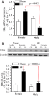
Figure 1.
Figure 1.
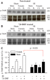
ERα mRNA and protein expressions in hippocampus at 3 d post-HI. ERα+/+ mice were subjected to either sham operation or HI. Three days, later the male and female hippocampi were probed for the quantification of ERα mRNA and ERα protein expressions. A, Hippocampal ERα mRNA expression was quantified 3 d post-HI using qPCR. Data is IL expression relative to sham-operated female, adjusted for CL ± SEM, n = 5–6. n=number of pups. B, Top, Representative blot of ERα protein expression in the IL and CL neonatal hippocampi from male and female mice subjected to either sham operation or HI. Blot was probed for β-actin as a loading control. Bottom, Summary figure of the ERα/β-actin ratio in male and female hippocampi of sham and HI mice. Data are mean IL adjusted for CL ± SEM, n = 3–4. n = number of blots per group (2 pups per group).
Figure 2.
Figure 2.
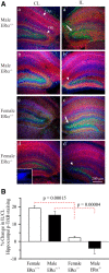
Effect of HI and 7,8-DHF on hippocampal TrkB phosphorylation in ERα+/+ mice. ERα+/+ mice were subjected to either sham operation or HI, with or without 7,8-DHF treatment. Three days later, the male and female hippocampi from the CL and IL sides were probed for phosphorylated TrkB (p-TrkB) expression, full-length TrkB (f-TrkB), truncated TrkB (t-TrkB), immature TrkB (i-TrkB), or degraded TrkB (d-TrkB). A, Representative blots of p-TrkB, and f-TrkB, t-TrkB, i-TrkB in the neonatal hippocampi of sham and HI ERα+/+ mice without 7,8-DHF treatment. B, Representative blots of p-TrkB, and f-TrkB, t-TrkB, i-TrkB in the neonatal hippocampi of sham and HI ERα+/+ mice with 7,8-DHF treatment. C, Summary figure of the p-TrkB/f-TrkB ratio in female and male hippocampi of sham and HI ERα+/+ mice with and without 7,8-DHF treatment. Data are mean IL adjusted for CL ± SEM, n = 3–4. n = number of blots per group (2 pups per group).
Figure 3.
Figure 3.
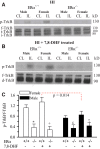
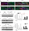
Immunohistological staining of p-TrkBY515 in ERα+/+ and ERα−/− mice 3 d post- HI. A, Changes in MAP2 and p-TrkBY515 immunoexpression in the CL and IL hippocampi of ERα+/+ and ERα−/− P9 male and female mice were examined 3 d post-HI. MAP2 (red), p-TrkBY515 (green), and DAPI (blue). Arrow, Increased p-TrkBY515 staining. Arrowhead, Decreased p-TrkBY515 in ERα−/− hippocampi. Inset, Primary antibody control. Scale bar, 200 μm. B, Summary figure showing the percent change in IL/CL ratio of hippocampal mean p-TrkBY515 fluorescent intensities in ERα+/+ and ERα−/− male and female mice hippocampi 3 d post-HI. IL/CL ratio of p-TrkBY515 obtained from uninjured naïve mouse hippocampus was assumed to be one. Three hippocampal slices per brain were analyzed. Data are mean ± SEM. n = 8 for ERα+/+ and n=3 for ERα−/−. n = number of pups.
Figure 4.
Figure 4.
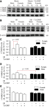
Effect of HI and 7,8-DHF on hippocampal p-TrkB in ERα+/+ and ERα−/− mice. ERα+/+ and ERα−/− mice were subjected to HI with and without 7,8-DHF therapy. Three days later, the male and female hippocampi from the CL and IL sides were probed for p-TrkBY705 expression, f-TrkB, t-TrkB, and i-TrkB. A, Representative blots of hippocampal p-TrkB (top), f-TrkB, t-TrkB and i-TrkB in ERα+/+ and ERα−/− mice without 7,8-DHF therapy 3 d post-HI. B, Representative blots of hippocampal p-TrkB (top), f-TrkB, d-TrkB (t-TrkB and i-TrkB) in ERα+/+ and ERα−/− mice with 7,8-DHF therapy 3 d post-HI. C, Summary figure of the hippocampal p-TrkB/f-TrkB ratio in ERα+/+ and ERα−/− mice with and without 7,8-DHF therapy 3 d post-HI. Data are mean IL adjusted for CL ± SEM, n = 3. n = number blots per group. *p = 0.018p, +p= 0.035q, εp= 0.334r, τp = 0.0488s versus ERα+/+.
Figure 5.
Figure 5.
Effect of HI and 7,8-DHF on hippocampal TrkB subtypes in ERα+/+ mice. The hippocampi of male and female ERα+/+ mice were probed for full-length TrkB (f-TrkB), truncated TrkB (t-TrkB), immature TrkB (i-TrkB), and β-actin. A, Representative blots of f-TrkB, t-TrkB, i-TrkB, and β-actin from sham-operated male and female neonatal hippocampi with and without 7,8-DHF therapy. B, Representative blots of f-TrkB, t-TrkB, i-TrkB and β-actin from male and female neonatal hippocampi with and without 7,8-DHF therapy 3 d post-HI. C, Summary figure of the f-TrkB/β-actin ratio in male and female hippocampi with and without 7,8-DHF therapy 3 d post-sham operation or post-HI. Data are mean ± SEM, n = 3–4. n=number of blots per group. D, Summary figure of the t-TrkB/β-actin ratio in male and female hippocampi with and without 7,8-DHF therapy 3 d post-sham operation or post-HI. Data are mean ± SEM, n = 3-4. n=number of blots per group. E, Summary figure of the i-TrkB/β-actin ratio in male and female hippocampi with and without 7,8-DHF therapy 3 d post-sham surgery or post-HI. Data are mean IL adjusted for CL ± SEM, n = 3–4. n = number of blots per group.
Figure 6.
Figure 6.
BDNF gene expression are sexually differentiated in P9 neonatal hippocampi 3 d post-HI. BDNF mRNA expression was quantified with qPCR in the hippocampi of male and female mice 3 d after sham operation or HI in ERα+/+ mice. Data is mean IL expression relative to sham female, adjusted for CL ± SEM, n = 5–6. n= number of pups.
Figure 7.
Figure 7.
Hippocampal p-srcY418 is sexually differentiated and ERα-dependent post-HI. A, Changes in the NeuN (red) and p-srcY418 (green) immunoexpression in the CL and IL dentate gyrus of ERα+/+ and ERα−/− P9 mice examined at 3 d post-HI. Left two columns = male; right two columns = female; top row = ERα+/+; bottom row = ERα−/−. Arrow, Increased p-srcY418 staining. Arrowhead, Unchanged or decrease p-srcY418 in IL hippocampi. Scale bar, 200 μm B, Representative blots of p-srcY418 and t-src from ERα+/+ male and female neonatal hippocampi. Mice were subjected to sham operation with and without 7,8-DHF therapy and the brains harvested 3 d later. C, Representative blots of p-srcY418 and t-src from ERα+/+ and ERα−/− male and female neonatal hippocampi. Mice were subjected to HI and the brains harvested 3 d later. D, Representative blots of p-srcY418 and t-src from ERα+/+ and ERα−/− male and female neonatal hippocampi. Mice were subjected to HI with 7,8-DHF therapy and the brains harvested 3 d later. E, Summary figure of the IL p-srcY418/t-src ratio adjusted for CL in male and female hippocampi 3 d post-sham operation or post- HI with and without 7,8-DHF therapy. Data are mean IL adjusted for CL ± SEM, n = 3–5. n = number of blots per group (2 hippocampi per group). F, Summary figure of the p-srcY418/t-src ratio in ERα+/+ and ERα−/−, male and female hippocampi 3 d post- HI with and without 7,8-DHF therapy. Data are mean IL adjusted for CL ± SEM, n = 3–5. n = number of blots per group. *p = 0.002t, +p= 0.001u , εp= 0.006v, τ p = 0.007w versus ERα+/+.
Figure 8.
Figure 8.
TrkB agonist treatment decreases hippocampal apoptosis in ERα+/+ female mice but has no effect in ERα −/− female mice. A, Brain slices from female P9 mice were stained for c-caspase-3 (green), NeuN (red), and DAPI (blue) at 24 h post-HI. Left two columns = female ERα+/+; middle two columns = female ERα+/+ with 7,8-DHF therapy; left two columns = female ERα−/−. Arrow head, Increased c-caspase-3 staining in ERα+/+ IL dentate gyrus. Arrow, Augmented levels of c-caspase-3 staining in ERα−/− IL dentate gyrus. Inset, Antibody control. Scale bar, 50 μm. B, C, Representative blots of c-caspase-3 in male and female neonatal hippocampi from ERα+/+ and ERα−/−with and without 7,8-DHF therapy 24 h post-HI. Blots were probed for β-actin as a loading control. D, Summary figure of the IL c-caspase-3/β-actin ratio corrected for CL in ERα+/+ and ERα−/− male and female hippocampi with and without 7,8-DHF therapy. Data are mean IL corrected for CL ± SEM, n = 3, n = number of blots per group (2 hippocampi per group).
Figure 9.
Figure 9.
Crosstalk between the ERα and TrkB post-HI. (1) HI induces ERα expression in neonatal hippocampal cells, which are thereafter activated by circulating or neural-derived estradiol (E2). (2) ERα signaling prompts activation of SFK, and (3) SFK enhance phosphorylation and hence, enhance activation of TrkB receptors in response to (4) TrkB receptor ligands resulting in (5) decreased apoptosis and increased neuronal survival.

Similar articles

Cited by

References

    1. Almli CR, Levy TJ, Han BH, Shah AR, Gidday JM, Holtzman DM (2000) BDNF protects against spatial memory deficits following neonatal hypoxia-ischemia. Exp Neurol 166:99–114. 10.1006/exnr.2000.7492 - DOI - PubMed
    1. Arpino G, Wiechmann L, Osborne CK, Schiff R (2008) Crosstalk between the estrogen receptor and the HER tyrosine kinase receptor family: molecular mechanism and clinical implications for endocrine therapy resistance. Endocr Rev 29:217–233. 10.1210/er.2006-0045 - DOI - PMC - PubMed
    1. Barletta F, Wong CW, McNally C, Komm BS, Katzenellenbogen B, Cheskis BJ (2004) Characterization of the interactions of estrogen receptor and MNAR in the activation of cSrc. Mol Endocrinol 18:1096–1108. 10.1210/me.2003-0335 - DOI - PubMed
    1. Bothwell M (2014) NGF, BDNF, NT3, and NT4. Handb Exp Pharmacol 220:3–15. 10.1007/978-3-642-45106-5_1 - DOI - PubMed
    1. Cazorla M, Arrang JM, Prémont J (2011) Pharmacological characterization of six trkB antibodies reveals a novel class of functional agents for the study of the BDNF receptor. Br J Pharmacol 162:947–960. 10.1111/j.1476-5381.2010.01094.x - DOI - PMC - PubMed

Publication types

LinkOut - more resources